Blog Page

Frozen Shoulder: Symptoms, Causes and Treatments

Doctors share why you may not want to wait out the discomfort of this middle-age malady

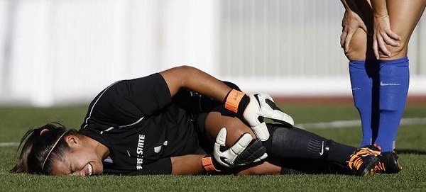
It can develop after an injury or out of the blue: One day you wake up with intense pain in your shoulder, and over the next few weeks the joint grows so stiff that your range of motion becomes severely compromised.

Known as frozen shoulder (aka, adhesive capsulitis), this mysterious condition usually occurs between the ages of 40 and 65 and affects more women than men. “It’s an interesting process, and it only happens to shoulders; we don’t see frozen knees or frozen elbows,” says Brian Feeley, M.D., a professor of orthopedic surgery at the University of California, San Francisco.

The problem arises where the head of the humerus, or upper arm bone, fits into a shallow socket in the shoulder blade — more specifically, in the shoulder capsule, which is the strong connective tissue surrounding the joint. “The name adhesive capsulitis says a lot because the [shoulder] capsule is the problem — there’s inflammation of the sac the shoulder lives in, and adhesive means it gets stuck after a while,” explains Beth Shubin Stein, M.D., an associate attending orthopedic surgeon and codirector of the Women’s Sports Medicine Center at the Hospital for Special Surgery in New York City.

The three stages of a frozen shoulder

In the early phase, the shoulder simply hurts and the pain gradually worsens. The second stage is the frozen stage, when the shoulder loses range of motion and “the capsule becomes thicker from inflammation,” notes Shubin Stein. The third stage, called the thawing phase, brings a gradual improvement to the shoulder’s range of motion. How long someone will spend in each stage is unpredictable and varies significantly from one person to the next, but the condition can take six months to two years or longer to resolve.

The exact causes of frozen shoulder remain a mystery, which is why it’s often referred to as idiopathic (meaning, of unknown cause). What is known is that it likely involves an element of inflammation, which triggers the release of chemicals that irritate the joint, causing pain, explains Jon J.P. Warner, M.D., cochief of the MGH Shoulder Service in Boston. “That, in turn, causes the release of other chemicals that cause stiffness or freezing and scarring.” More often than not, frozen shoulder affects the non-dominant arm, and if someone develops idiopathic frozen shoulder in one shoulder, she’s at higher risk for getting it on the other side later, Warner says.

Your risk factors for feeling the freeze

Research has found that people with diabetes are five times more likely to develop adhesive capsulitis; those with thyroid disorders have a nearly three times higher risk of developing the condition. More common in women overall, frozen shoulder can also happen after breast cancer treatment or heart surgery, Warner explains. And there appears to be a genetic predisposition to frozen shoulder, meaning that if a first-degree relative had it, you may have a higher risk.

In 2013, Stacey Marnell, a social worker and school counselor who lives in Takoma Park, Maryland, experienced pain and gradual stiffening in her right shoulder, which progressed to the point where she couldn’t lift her right arm above shoulder level. “My doctor said I was a triple threat for frozen shoulder because I have type 1 diabetes, I take thyroid medications and I’m a petite woman,” says Marnell, 67. “I felt desperate because it was limiting everything I could do — I couldn’t do zippers or buttons on my back, open a window or dry my hair.”

Addressing the problem

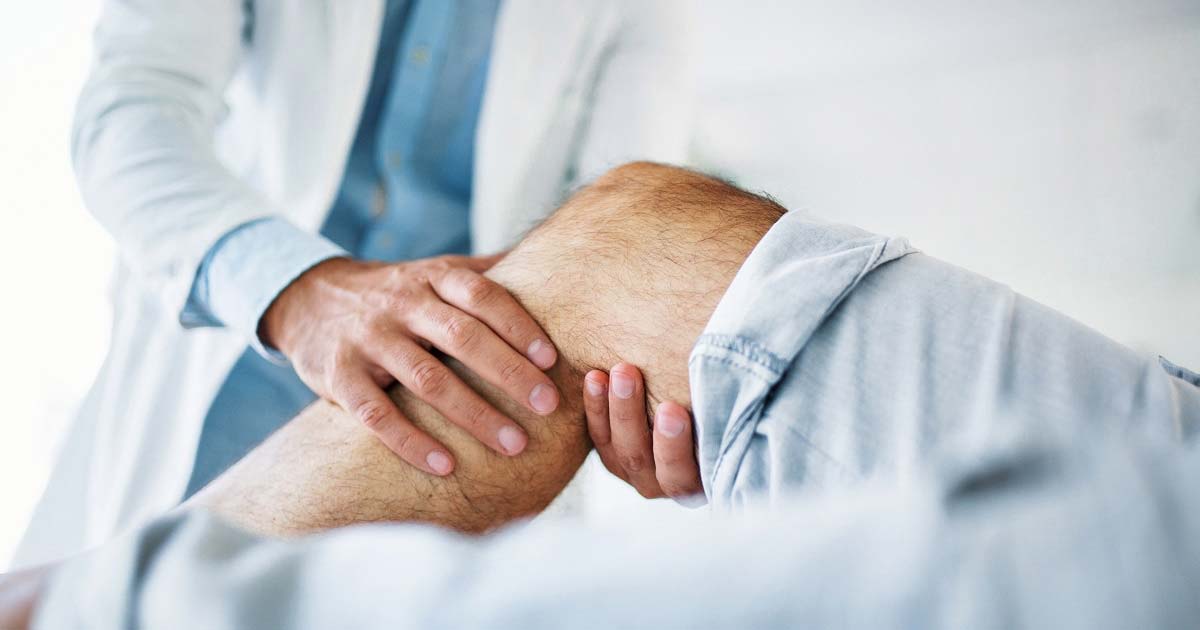
A doctor can diagnose frozen shoulder based on a patient’s description of when symptoms began and when the pain occurs, as well as by doing a physical exam. Identifying loss of active motion (voluntarily being able to move the affected arm and shoulder in certain directions) and loss of passive motion (when the physician tries unsuccessfully to move the arm/shoulder in different directions) is key to making the proper diagnosis, Warner says. An X-ray is taken to rule out the possibility of arthritis in the shoulder causing the limited motion.

“For a long time the treatment was benign neglect, because if you don’t do anything, it will eventually go away,” Shubin Stein says. “But it’s a miserable path for those two years.” And sometimes the recovery process takes longer and can leave the person with permanent losses to their range of motion, she adds.

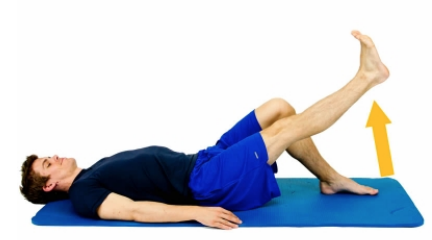
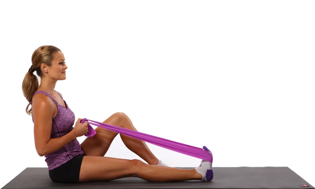
These days, more cases are addressed, though “treatment is complicated because a lot of things work, but nothing works perfectly,” Feeley says. The goals are to “increase range of motion in the affected shoulder and to decrease the cascade of inflammation and scarring in the capsule.” The general approach to treatment involves anti-inflammatory medications — taken orally or given as a cortisone injection into the shoulder joint if frozen shoulder is in an early stage — and physical therapy. With PT, specific exercises are performed to gently stretch the tissues in the shoulder and improve range of motion — through external rotation and things like crossover arm stretches, for example.

Is There a COVID-19 Connection to Frozen Shoulder?  

Following anecdotes about middle-aged COVID-19 survivors subsequently developing frozen shoulder, researchers explored the possible link in a study that was published in the July 2021 issue of the Journal of Shoulder and Elbow Surgery. They confirmed what they call a possible association between the disease and the shoulder condition. Interestingly, those who had asymptomatic or mild COVID-19 infection had higher rates of frozen shoulder.

The researchers theorize that the link between the two may be due to COVID-19’s direct infection of the cells outside the respiratory tract, as well as from “a cytokine storm and a systemic inflammation that may impact nearly every organ system, including the musculoskeletal tissues.”

Though he hasn’t seen this pattern in his practice, orthopedic surgeon Feeley says, “It makes sense that some people who develop COVID, which causes an intense immune reaction, are going to end up with frozen shoulder. There could be a relationship [with the virus], but it hasn’t been clearly defined.” Experts have also suggested that factors such as being sedentary while sick during COVID-19 could play a role.

Maria Leonard Olsen’s frozen shoulder started as mild pain in her right shoulder. From there it gradually became more and more difficult to lift her arm as the discomfort worsened. “It’s a weird, throbbing pain; it’s so intense, and then it just stops,” says Olsen, 58, a lawyer and writer in Bethesda, Maryland. This past July, she saw an orthopedic surgeon, who made the diagnosis, gave her a cortisone shot and sent her for PT twice a week. “It’s about 50 percent better now,” she says, both in terms of movement and pain.

If the more conservative treatments don’t help, surgery may be an option during stage 2 of frozen shoulder. With a procedure called arthroscopic capsular release, an orthopedic surgeon releases the stiffened joint capsule and removes adhesions (scar tissue), Warner explains. Surgery is usually followed by intense PT to help the patient maintain the restored range of motion.

To treat her (right) frozen shoulder, Marnell went for physical therapy and dry needling (which involves inserting super thin needles into muscles, tendons, ligaments or other tissues to increase range of motion and reduce pain). The combination helped a bit but not completely. Meanwhile, her left shoulder started stiffening up, and it didn’t respond to stretching, PT, massage or acupuncture. (While there’s little scientific evidence that acupuncture or dry needling helps, these techniques may be helpful as adjunctive therapies, Shubin Stein says.) In 2015, Marnell had arthroscopic surgery on her left shoulder to break up the frozen tissue, followed by intense PT. These days she has nearly full mobility in both shoulders.

To maximize recovery from frozen shoulder, early diagnosis is important, experts say. If shoulder pain is waking you up at night, if you’re losing range of motion (especially when rotating your shoulder inward), or if fast motions with your upper arm hurt more than slow motions, see a doctor. “Early intervention and diligent treatment make this a much quicker process if we catch it before scarring occurs,” Shubin Stein says.

Read the article on AARP site.

adminFrozen Shoulder: Symptoms, Causes and Treatments
read more

Dislocate Your Knee? What to Know About Patellofemoral Instability

Smooth knee movement can be affected by this condition. Find out more about what it means and how to treat it.

For most people, the kneecap, or patella, glides nicely within a V-shaped groove at the end of the thigh bone called the trochlea. As you bend and straighten the knee, the patella moves up and down and is stable within the trochlea, allowing you to stand, sit, walk and run without a problem.

Unfortunately, for others, things can go awry. “Sometimes we think of the patella as the train, and the notch it sits in, called the trochlear groove, as the track,” says Theresa Chiaia, PT, DPT, a physical therapist at HSS. “The issue for patients who dislocate their patella is that the train comes off the track, leading to the knee giving way, as well as pain and swelling.”

In a complete dislocation, the patella comes out of the track completely. Some people may experience more subtle issues with instability, which is known as subluxation, or partial dislocation. A partial dislocation also results in pain, swelling, popping or cracking sensations or stiffness, but these are much less severe, and the recovery is usually days instead of weeks or months.

Whether a person has a partial or a complete dislocation, the soft tissues of the knee, including the ligaments and cartilage, are often injured. If the ligament that keeps the patella in place, called the medial patellofemoral ligament (MFPL), is stretched or torn, it increases the chance that dislocation will continue to happen.

How do you know you if have dislocated your patella?

If you have a partial dislocation, the signs may be subtle. You may have pain around the kneecap, or it may feel like the knee is popping out and popping back in.

A dislocation is usually more obvious. The affected knee will buckle and be unable to support your weight, and you may have significant swelling, pain and stiffness. The kneecap may pop itself back in, or you may need help from someone else to get it back in place. Even if the kneecap shifts back into place on its own, pain and swelling can remain.

Who is at risk for a dislocated patella?

While these issues can happen in men and women, young and old, they happen most frequently to young women in their teens or early 20s. People who have patella dislocations usually have some physical risk factors, such as a notch (trochlea) that is not shaped correctly; significant laxity, or looseness, in their soft tissues; or a kneecap that sits too high on the thighbone, all of which can be hard for someone to know without being told by a doctor.

How is a dislocated patella diagnosed?

After listening to the story of how the injury happened, your doctor will perform a physical exam. The exam will begin with you seated and moving your knee. Then you’ll probably lie down and your doctor will bend your knee slightly and guide your kneecap outward. This feels uncomfortable and potentially painful for people with patellar instability. Your doctor may take measurements to see how the bones are aligned, and he or she may watch you walk as well.

Patients typically will have an X-ray to see how the kneecap fits in the groove, though if the kneecap has already returned to the correct position, the X-ray may look normal. This is why the physical exam is so important. You may also need an MRI to look at the ligaments and cartilage to check for damage, which to some degree is always seen after a dislocation. However, if that damage is minimal and it is the first time it has happened, then you may be treated without surgery.

Is it possible to dislocate your patella more than once?

People over the age of 25 who dislocate their patella for the first time have a fairly low chance of it happening again if they are treated appropriately with a brace, crutches and physical therapy. These people often don’t need surgery. People who are under 25 and who have a shallow groove have a much higher chance of the problem continuing to happen. These patients may need surgery. “That’s why it’s so important to manage these issues right away,” says Chiaia. “If you stop the kneecap from dislocating, you can stop further damage from happening.”

How do you treat a dislocated patella?

If the kneecap is dislocated and hasn’t returned to its proper place on its own, it needs to be put back where it belongs. Your doctor will apply gentle pressure to push it back, a process called reduction. Afterwards, rest, a knee brace and crutches will help the swelling to go down and allow the knee to heal. If the knee is very swollen, draining the knee of fluid can help reduce the discomfort. After a week or two of rest, physical therapy can help strengthen the muscles around the kneecap to help keep it aligned in the trochlear groove, and help the knee get back to a normal range of motion.

If an MRI shows that bone or cartilage has been damaged on the thigh bone or on the underside of the kneecap, arthroscopic surgery may be necessary. Both of these issues can cause locking, buckling or additional pain in the knee. In people who have had more than one dislocation, surgery can recreate the ligament that was torn. Sometimes in addition to ligament surgery, the doctor will recommend realigning the bone in a procedure called a tibial tubercle osteotomy to help keep the kneecap on track. The success rate of surgery for patellar instability is very high. More than 95% of patients have no more dislocations, and more than 85% return to sports they played before their injury at the same or a higher level.

Read the article on HHS site.

adminDislocate Your Knee? What to Know About Patellofemoral Instability
read more

Patellofemoral Syndrome: Covid-19 Edition

Over the past few months, people have been getting creative on how to stay active and healthy during the covid shut down.  For many, running has become an outlet for both mental and physical health.  

Patellofemoral syndrome, commonly referred to as runner’s knee, is an exceeding common condition and we’ve posted about it before.  However, over the past few weeks I’ve noticed a bit of an increase for this condition in our practice.  With gyms closed, it is difficult to stick with a well-rounded routine, balancing both cardio and strengthening.  Without access to gym equipment or a body sculpting class, many are forgoing the strengthening side of the equation altogether, which is contributing to the development of patellofemoral syndrome.  

Whether you are a new runner, or a veteran, it is imperative to supplement running with a strengthening program that focuses on core, gluts, and quads.  Below is a home exercise program we typically recommend to patients.  With the exception of the leg press exercise (which you can skip for now), no equipment is required.  Add bands or light weights as you advance. 

adminPatellofemoral Syndrome: Covid-19 Edition
read more

Managing your ACL injury during COVID-19

Hospitals throughout the country have canceled elective surgery in order to decrease the spread of COVID-19 to patients and healthcare workers and to concentrate their efforts on emergency care. We recognize the impact this has had on our patients. They may have an ACL injury earlier in the year and are hoping to be ready for next season to compete in cutting and pivoting sports or they may rely heavily on a stable knee to perform their jobs. Unfortunately this situation has forced patients to have to wait out their injuries at home until facilities can be reopened to treat their injuries. While things may seem bleak at the moment there are things you can do at home now to help prepare yourself for upcoming ACL surgery. A good at home physical therapy prehabilitation program can help you stay focused and improve your readiness for surgery. Here are some of the basic goals for ACL prehabilitation:

  • Decrease pain   
  • Minimize swelling
  • Maximize knee range of motion(knee bending & straightening)
  • Normalize gait (way we walk)

This Home Exercise Program developed by HSS PT will take you through some exercises that will help you to work toward the above goals with the aim of getting you conditioned for your surgery. If you have been working with a physical therapist already this is a good time to discuss our prehab program with them and stay in touch with them via phone, email or virtual channels as you progress. We recognize that given work and family constraints it may be difficult to find the time but if you can dedicate a few hours per week it can be very beneficial. The current state of our daily lives has been immensely impacted by the crisis but your health and safety is our #1 priority. During these uncertain times what we do know is that healthy habits and movement can have a big impact on your state of mind. Be well and be safe!

adminManaging your ACL injury during COVID-19
read more

Patella dislocations- what we know and what we don’t

Having a patella dislocation can be a very scary and stressful injury for patients. While any injury can be difficult to deal with, in my experience patella dislocations in particular can be anxiety producing for patients because there is the fear of the unknown – the fear that their patella will dislocate again or the fear that it will happen to their other knee, and how this will affect their ability to continue playing sports or go to school or work.  It also conjures up thoughts of needing surgery. The injury can happen from a contact injury, such as colliding with another player during a soccer game, or from a sudden twist or turn. The injury happens very quickly and in some instances the individual may fall to the floor and they may be unable to walk. Their knee may get very big and swollen, and they may end up in the emergency room in order to get their patella relocated. Then they are typically sent home with a brace and instructed to see an orthopedist who specializes in patella injuries like myself. This may be the individual’s first time having such an injury or they may have had recurrent episodes of the patella “shifting” but not dislocating in the past, or this may be the tenth time their patella has dislocated.  Regardless of the circumstances, this injury can take a psychological toll on patients and understandably so.

I have dedicated my career to understanding the complexities and intricacies of the patella and there is still a lot we have to learn. I have authored books and done extensive research on the subject of patella instability and pain and have helped develop systematic approaches to the surgical management of these injuries including medial patellofemoral ligament reconstruction (MPFL). I have performed countless surgeries to correct these injuries and have had excellent results and success. Despite all my efforts there is still a lot we don’t know when it comes to selecting the right patients for surgery. For example, a 16-year-old competitive soccer player who comes into my office with her parents after her first patella dislocation during a soccer game. They are devastated by her injury and they may want to have the surgery done as soon as possible to maximize recovery time so as not to miss the following season. The anxiety and tensions are obviously very high, and we have an extensive conversation about patella instability including why the injury likely happened, what the risks are that this will happen again and what I feel is the best approach to managing her injury. Then comes the most difficult part of the conversation, which is that we know that MPFL reconstruction is a safe and effective procedure but what we don’t know for certain yet is whether or not a first-time dislocator should have surgery. The standard of care currently is that non-surgical treatment is the best option for first-time dislocations unless there are other related injuries such as a displaced piece of cartilage in the knee. Explaining to a 16-year-old competitive soccer player and her family that she has a high risk of another dislocation but that she should be treated without surgery is a very difficult conversation to have. This is why I have dedicated years to my current research studies to be able to make a more informed decision with my patients. I am leading a study that aims to answer whether a young first-time dislocator should be treated with surgery called PAPI (Pediatric and Adolescent Patellar Instability). This is a multi-center study in which patients who are selected randomly to either be treated with surgery or without surgery after their first patellar dislocation. Another study that I am leading called JUPITER is a large multi-center patient registry on patellar instability and is the first of its kind in the US. We currently have 28 orthopedic surgeons at 13 centers across the country contributing to JUPITER, and to date have enrolled over 1,200 patients with patellar instability. In the not too distant future, the hope is that the results of these studies will give us a clearer picture of how we should be managing first-time dislocators and patella instability in general, and will allow me to be able to have a more informed conversation with my patients and their families and help to alleviate some of their anxiety and concerns.

adminPatella dislocations- what we know and what we don’t
read more

Spotlight Patella: Interesting facts about this simple yet complex structure in our knee.

Did you know ostriches are born with four kneecaps (patella)? The evolutionary reason for this is still not fully understood but theories include that it allows this heavy fast bird to straighten its knee faster thereby helping it to run faster. I have dedicated so much of my career to issues of the human patella. Many issues can arise from this small but fascinating bone. See below to learn some fun facts about this simple yet complex structure in our knees. 

Ostrich knee X-ray
  • The patella is a roughly triangular shaped bone called a sesamoid bone from the Latin sesamun “sesame seed”. It is the largest of this type of bone in our body as compared to the tiny sesamoid bones in say our foot.
  • Babies are born with a patella made of cartilage instead of bone partly due to its flexibility for the birthing process
  • The patella slowly turns from cartilage to bone between the ages of 2-6 years old
  • Some people’s patella bone never fuses and so they may have two separate bones know as a bipartite patella. Rare genetic disorders cause some people to not develop a patella bone.
  • “The knee bone is connected to the thigh bone” – this is sort of true as the quadriceps tendon in your thigh attaches to the upper part of your patella and patella tendon below your patella attaches to your shin bone.
  • The patella sees an average of 2.5-3.5 times your body weight with stairs and up to 7-8 times your body weight with deep squat.
  • The patella is vital to protect the inner structures of your knee acting as a shield. The patella increases the leverage of the knee extensor (straightening) muscles, so they don’t need as much force to straighten the knee.
  • For such a small bone it is prone to injury including fractures, dislocations, patellofemoral syndrome and arthritis. Some people even have the patella removed due to severe injury or arthritis.

These are just some of the interesting facts about the patella and demonstrate how intricate and fascinating a structure it is in our bodies. Given that injuries to this bone and surrounding tendons, ligaments and joints are so common, it is important to properly manage these injuries to prevent damage to the surrounding structures as they are all interconnected. If you are experiencing issues of the patella or patellofemoral joint please visit the patellofemoral disorder section of my website to learn more.

 By Dr. Beth Shubin Stein

Spotlight Patella:  Interesting facts about this simple yet complex structure in our knee.


adminSpotlight Patella: Interesting facts about this simple yet complex structure in our knee.
read more

I am double jointed. Does this mean I have more joints that other people?

The term double-jointed is a misnomer. The name implies that a person has twice the number of joints as normal and thus their joints allow for greater movement outside of the normal range of motion for that joint. What this term actually refers to is that a person has greater mobility or flexibility of their actual joint and can move their joint beyond its normal end range without experiencing pain and discomfort. Hypermobile joints tend to be inherited. It occurs in about 10-25% of the general population.

Most people don’t experience any symptoms and don’t need any intervention, but a small percentage of patients will have hypermobility syndrome where their unstable joints can lead to other conditions. Some may experience frequent sprains or tendinitis of their joints and activity modification and physical therapy may be recommended to strengthen and stabilize their joints. In some rare cases, these joint issues may be related to a more serious underlying medical condition. In my practice, I often see patients who experience subluxations or dislocations especially of their knee cap (patella) or their shoulder joint due to hypermobility. A subluxation is when the bones of a joint partially move out of place and then relocate back in on their own (known as a partial dislocation) versus an actual dislocation where the bones move completely out of the joint and stay out until relocated. These injuries may occur from direct trauma to the joint like during sports or from a fall.

I routinely see another subset of patients in my practice who have abnormal joint anatomy where the ends of their bones in their joint are abnormally shaped. These patients can not only have laxity in their joints but also have anatomy that predisposes them to injury. These patients can experience a subluxation or dislocation event even without trauma such as from a sudden twist or movement of their joint. These patients require a thorough evaluation to determine what the best treatment is for them. It can range from pain medication and physical therapy to surgeries to stabilize the joint and prevent re-injury. Having hypermobile joints is not a problem for most people, but if pain and frequent injuries occur it is important to see an orthopedist who regularly deals with these issues. 

adminI am double jointed. Does this mean I have more joints that other people?
read more

My teammate had an ACL repair, why is a different ACL surgery being recommended for me?

Over the years in my practice, this has become a familiar question—many patients come to my clinic wondering about their options for anterior cruciate ligament (ACL) surgery. These patients range from someone who tripped and fell to recreational athletes who sustained an ACL tear skiing, to the amateur/professional athletes who sustained an ACL tear competing in high-level, pivot-heavy sports. My first response to my patients is that there is no one-size-fits-all approach to ACL surgery. I begin by explaining the difference between an ACL repair (where the ligament is sewn back to its origin) versus an ACL reconstruction (where a new ligament is constructed for them). We discuss that every case is unique and that there a multiple factors that go into determining the right type of surgical or non-surgical solution for their ACL injury, including:


  • Location and type of tear:
    Most ACL tears occur in the middle or mid-substance of the ACL, and can be partial, meaning part of the ligament is torn, or complete, where the ligaments is torn in half. Less frequently we see an avulsion type ACL injury, where the ACL “peels off” from its attachment to the bone. These injuries are all ACL tears, but the treatments are not all the same.
  • Chronicity of the tear:
    We want to know if this is a recent ACL injury, or if the ACL injury occurred months to years ago, as this affects healing patterns.
  • Age & activity level:
    The age, activity level, and future expectations of the patient are each critical factors in determining the right approach to ACL surgery. Is the patient low demand, meaning they want to get back to bike riding or yoga or are they high demand college athlete competing in cutting and pivoting sports. 
  • Surgical history:
    I am looking to determine what prior knee surgery the patient has had on the injured knee, as well as the other knee and joints. 
  • Imaging results:
    These results help us see if there is a fracture or break in the bone where the ACL attaches, a tear in the meniscus (shock absorber), damage to the cartilage (smooth glossy coating over the bones) and damage to other ligaments. These factors also affect the type of treatment that may be needed.   
  • Physical exam results
    This includes an evaluation in the office where we assess areas of pain and other stabilizing ligaments in your knee to see how stable your knee is. We also examine the anatomy of your knee to ensure proper alignment and mobility of your hips, knees and ankle joints.  

These are just some of the factors that go into making the appropriate treatment choice to heal your ACL injury. While your friend, colleague or teammate may have had a different approach and experience with their ACL injury treatment, it doesn’t mean yours will be the same. Having a detailed discussion with your doctor about your options and coming up with a plan that works best for you is ultimately the most important factor.

adminMy teammate had an ACL repair, why is a different ACL surgery being recommended for me?
read more

You Don’t Need to Run to Have Runner’s Knee

Beth Shubin Stein, MD, Associate Attending Orthopedic Surgeon

Sabrina Strickland, MD, Associate Attending Orthopedic Surgeon

HSS Patellofemoral Center

Do you feel pain in the front of your knees when going down stairs? Does knee pain interfere with your ability to walk, run, kneel, squat, or stand up comfortably? Has your knee ever “given out” on you? If you answered yes to any of these questions, you may be suffering from patellofemoral knee pain, arthritis, or instability. Anyone can suffer from these conditions, but they are more common in women than men.

Commonly known as “runner’s knee,” patellofemoral (“anterior knee”) pain can affect anyone. It can happen in people with improper alignment, where the kneecap does not slide smoothly in the groove or track of the femur (thighbone). This type of problem may run in families. Or it may result from injury to the knee (usually during adolescence or young adulthood).

Some people feel pain around or under the kneecap. Others feel instability and may experience a kneecap dislocation. Not everyone with patellofemoral problems needs surgery, but in some cases it is warranted to prevent more serious problems down the road. Here’s how to know what may lie ahead for you. (See a doctor to know for sure.)

If You Have Knee Pain

If you have knee pain without dislocation of the kneecap and a short course of modifying your activities does not resolve the discomfort, see a primary care sports medicine doctor or an orthopedic surgeon. You’ll likely have an x-ray of your knee to see how the kneecap is tracking in the groove of the femur. Physical therapy may help to strengthen the muscles around the knee that help keep the kneecap in place. If your pain persists despite physical therapy, you may need an MRI to look for cartilage damage under the kneecap.

If You Have Arthritis

If tests show there is cartilage damage under your kneecap (arthritis), you may continue physical therapy and/or receive injections to reduce inflammation and provide lubrication. Your doctor or physical therapist may advise you to change, reduce, or avoid certain activities that may aggravate your symptoms. Some patients with cartilage damage may have surgery with techniques that allow regrowth of the damaged cartilage or replace damaged bone with donor bone and cartilage. The surgeon may also correct any misalignments to prevent or minimize future cartilage damage. Patellofemoral cartilage surgery is typically reserved for younger patients (under age 35) or for older patients whose pain persists despite nonsurgical treatments. Some older patients may have partial knee replacement to create a new smoothly gliding joint.

If You Have Kneecap Instability

When the kneecap doesn’t track evenly in the groove of the thigh bone, it can slide out to the side (dislocate), causing your leg to give out under you. This instability is most common in adolescents and young adults, and it is also more common in females. But it can happen in anyone at any age with patellofemoral malalignment and trauma.

Doctors used to think that patients who had only one dislocation should always try nonsurgical treatment. However, much has been learned in the last five years indicating which patients with a first-time dislocation have the highest risk of recurrence. As a result, all patients who have a dislocation should see an orthopedic surgeon to determine if they are in that high-risk group and might benefit from early surgery.

If your kneecap dislocates a second time, you will need surgery to reduce the risk of arthritis from continued dislocations. During the procedure, the surgeon may rebuild the medial patellofemoral ligament — a “leash” that holds the kneecap in place, which often tears during dislocation and then heals in a stretched-out position. This can be done using your own or donor hamstring tissue; in some cases, you may require an additional bony surgery to fix problems with poor alignment. We are currently doing research to identify factors that increase the risk of a second dislocation among first-timers and potentially perform surgery earlier in those people to prevent subsequent dislocation.

If your kneecap has dislocated, it’s extremely important to have it checked out. Each time you experience a dislocation, there is likely to be damage to the cartilage that increases your risk of arthritis. See a sports medicine physician or orthopedic surgeon to find out what’s best for you.

adminYou Don’t Need to Run to Have Runner’s Knee
read more