

PATELLAR INSTABILITY
What is Patellar Instability
Patellofemoral Instability is when the kneecap (patella) either slips partially out of the track of the joint—this is called subluxation—or is completely dislocated. When this happens, the knee snaps outward, causing the MPFL (medial patellofemoral ligament) to tear or stretch. This usually occurs when playing a sport, and results in the buckling of the knee, followed by a fall.
The Medial Patellofemoral Ligament (MPFL) is: The ligament that attaches the inside part of the kneecap (or patella) to the long bone of the thigh, also called the femur.
People at risk for subluxation and dislocation include:
- Athletes who experience a traumatic dislocation while competing
- Young patients (commonly females) who are loose jointed
The risk factors of Patellofemoral Instability are:
- A shallow (or absent) groove on the trochlea or femur
- An abnormal lateral attachment of the patellar tendon on the tibia (shin)
- Knock knees
- High riding kneecap
What are the Symptoms of Patellar Instability
Signs of dislocation include:
- Significant swelling of the knee
- An “apprehension sign,” or anxious response to the doctor pushing the patella outward in an attempt to mimic the dislocation
Having an MRI after a kneecap dislocation reveals damage to the ligament (the MPFL), as well as bruises on inside of the patella, and on the outside of the femur. The MRI also helps your doctor evaluate the knee for evidence of cartilage injury, which is common after dislocations.
What are the Treatments for Patellar Instability
Non-Surgical Treatment
If your injury doesn’t require surgery, your knee will usually be placed in a brace for a few days to several weeks to allow any swelling and pain to subside. Your orthopedist may also drain fluid from the knee to reduce discomfort if there is considerable swelling.
Physical therapy is started within the first 1 to 2 weeks after the dislocation. Your sessions will generally continue for 2 to 3 months after a dislocation, and recovery can take as long as 4 to 5 months.
After your first dislocated kneecap, you have an increased risk of it happening again. Although the injured ligaments do heal during recovery, they usually do so in a stretched-out state, further contributing to the risk of another instability episode.
Surgical Treatments
What is the procedure?
This procedure consists of several small incisions around the knee to utilize a camera to visualize the inside of the knee. The surgeon will then debride the problem area or remove the loose body.
How long will I stay in the hospital?
This surgery is typically done as ambulatory surgery, meaning you will go home the same day of surgery.
What are the possible risks and complications of surgery?
As with any surgery there is a risk of DVT, nerve damage, and postoperative infection. Specific risks and complications include but aren’t limited to persistence of symptoms and post-op stiffness.
When can I drive?
You may not drive while taking pain medication. In addition, if it is your right knee that had surgery, you will not be able to drive for approximately 2-4 weeks after surgery.
When will I be back to all normal activities?
This typically is around 2-3 months after surgery. You will be allowed to begin biking without resistance once you have adequate range of motion and will begin this with your physical therapist, typically 2-3 weeks after surgery. Once adequate quad strength is demonstrated you will transition to the Elliptical, then to running, and then to more specific activity if desired. Please see Your Surgery and Physical Therapy for more information on this condition.
What is the medial patellofemoral ligament (MPFL)?
The MPFL is a ligament that stabilizes the patella (kneecap). It is disrupted when someone dislocates their patella. It helps to restrain your kneecap from displacing laterally.
How is the MPFL damaged?
The general mechanism behind MPFL injury is lateral patella dislocation. When the kneecap dislocates towards the outside, this stretches the ligament on the inside of the knee, which is trying to keep the kneecap in place. This can result in either a tear of the MPFL or a detachment of the ligament from the bone. Generally, this injury occurs with twisting or turning of the leg.
How will my MPFL be reconstructed?
This procedure is done through a small incision made at the inside portion of the knee. The injured ligament will be replaced with a graft, usually a hamstring tendon from the same leg or a cadaver allograft. The graft is attached to the patella via small absorbable screws that hold the graft in place.
Here is a nice animated video detailing how MPFL reconstruction surgery is performed including information about what to expect on the day on surgery.
How long will I stay in the hospital?
This surgery is typically done as ambulatory surgery, meaning you will go home the same day of surgery.
What are the possible risks and complications of surgery?
As with any surgery there is a risk of DVT, nerve damage, and postoperative infection. Specific risks and complications include but aren’t limited to re-tear, post-op stiffness, and arthritis.
When can I drive?
You may not drive while taking pain medication. In addition, if it is your right knee that had surgery, you will not be able to drive for approximately 6 weeks after surgery or until the brace is removed.
When can I resume jogging?
You will not resume jogging until cleared by your physician. This typically is around 4-6 months after surgery. You will be allowed to begin biking without resistance once you have adequate range of motion and will begin this with your physical therapist. Once adequate quad strength is demonstrated you will transition to the Elliptical, then running is the next step after that.
When can I return to my sport?
There are many factors in returning to sport after surgery. Most patients are able to return around 6 months after surgery. Please see Your Surgery and Physical Therapy for more information on this condition.
What makes me a candidate for this surgery?
You are a candidate if you have patellar instability (kneecap dislocation) and focal cartilage injury. This is evaluated through your past history and the use of MRI and XRAY.
What is the medial patellofemoral ligament (MPFL) and how is it damaged?
The MPFL is a ligament that stabilizes the patella (kneecap) and keeps it from displacing laterally. It is disrupted when someone dislocates his/her patella. When the kneecap dislocates towards the outside, this stretches the ligament on the inside of the knee, which is trying to keep the kneecap in place. This can result in either a tear of the MPFL or a detachment of the ligament from the bone.
What is cartilage, why is it important, and how is it injured?
Cartilage is the shiny, smooth coating at the end of bones. It protects the bone and allows the bones to move smoothly and efficiently. Damaged cartilage is known as arthritis. When cartilage thins, or has a piece missing, it puts more stress on the bone and causes pain. The mechanism of injury for a cartilage defect is usually related to trauma, such as a dislocation that causes the cartilage to scrape along bone, or chronic friction from a maligned knee. The body cannot regenerate this type of cartilage.
What does the surgery entail?
This procedure is done through two small open incisions made at the inside portion of the knee and midline along the patella; and through two small arthroscopic incisions. The injured ligament will be replaced with a graft, usually a hamstring tendon from the same leg or a cadaver allograft. The graft is attached to the patella via small absorbable screws that hold the graft in place. The cartilage defect is debrided and restored using one of the methods described below. The incision is then closed with absorbable sutures and Dermabond, a surgical glue.
How will my cartilage be repaired?
This depends on many factors including your age as well as the size and location of the cartilage defect. The surgeon will chose from one of the following procedures:
– Microfracture
- Procedure: Using arthroscopy, the surgeon will first remove any defective and damaged cartilage tissue from the knee joint. She will then create small holes at the site of your lesion to allow bleeding which will stimulate healing. The new tissue that grows is a hybrid of fibrocartilage and another type of cartilage that is similar to that originally in the joint. Although it is not exactly the same, this new type of cartilage is shown to be durable and to function similarly to the original articular cartilage. This procedure can be used on very small lesions successfully.
– DeNovo
- Procedure: The surgeon will first remove any defective and damaged cartilage tissue from the knee joint. She will then patch the cartilage defect with the DeNovo cartilage cells. This consists of juvenile cartilage which is able to rejuvenate and grow.
- You may require follow-up MRIs at 3 mo, 6 mo, 12 mo, and 24 mo after surgery to follow the growth and maturation of the new cartilage.
– OCA (Osteochondral Allograft)
- Procedure: The surgeon will first remove any defective and damaged cartilage tissue from the knee joint. She will then patch the cartilage defect with a plug of allograft donor cartilage and bone. This does not rejuvenate, but is press fit into the bone. The body then grows into the new plug and it becomes your own.
– MACI (autologous cultured chondrocytes on porcine collagen membrane)
- Procedure: This is a staged surgery. Stage 1- Using arthroscopy, the surgeon will first remove any defective and damaged cartilage tissue from the knee joint. Healthy cartilage is biopsied and sent to a lab where the cartilage cells will proliferate. Stage 2- About 4-6 weeks later, the defect will be patched with your new cartilage cells.
How long will I stay in the hospital?
This surgery is typically done as ambulatory surgery, meaning you will go home the same day of surgery.
What are the possible risks and complications of surgery?
As with any surgery there is a risk of DVT, nerve damage, and postoperative infection. Specific risks and complications include but aren’t limited to failure to heal, fracture, and hardware complications.
When can I drive?
You may not drive while taking pain medication. In addition, if it is your right knee that had surgery, you will not be able to drive for approximately 6 weeks after surgery or until the brace is removed.
When can I resume jogging?
You will not resume jogging until cleared by your physician. This typically is around 5-6 months after surgery. You will be allowed to begin biking without resistance once you have adequate range of motion and will begin this with your physical therapist. Once adequate quad strength is demonstrated you will transition to the Elliptical, then running is the next step after that.
When can I return to my sport?
There are many factors in returning to sport after surgery. Most patients are able to return around 7-10 months after surgery. Please see Your Surgery and Physical Therapy for more information on this condition.
Am I a candidate for PFJR + MPFL?
You are a candidate if you have patellar instability (kneecap dislocation) and front knee pain with examination findings that point towards kneecap arthritis (patellofemoral osteoarthritis). This is evaluated through the use of MRI and XRAY.
What is osteoarthritis and how does it occur?
Cartilage is the shiny, smooth coating at the end of bones. It protects the bone and allows the bones to move smoothly and efficiently. When this cartilage thins or becomes damaged, it’s called osteoarthritis. It can be caused by a number of factors including genetics and injury.
What is the medial patellofemoral ligament (MPFL) and how is it damaged?
The MPFL is a ligament that stabilizes the patella (kneecap) and keeps it from displacing laterally. It is disrupted when someone dislocates his/her patella. When the kneecap dislocates towards the outside, this stretches the ligament on the inside of the knee, which is trying to keep the kneecap in place. This can result in either a tear of the MPFL or a detachment of the ligament from the bone. Additionally, when the kneecap goes out of place, and then back in, it knocks on the lateral femoral condyle (part of the thigh bone), which can often result in an injury to the cartilage and contribute to arthritis.
What does the surgery entail?
A vertical incision is made in the front of the knee. Focusing on the patellofemoral compartment, the damaged bone and cartilage in the knee joint are removed and the worn ends of the bone are shaped to fit the implant, which is inserted and attached to the bones with cement. The exact type of implants used is called Zimmer. The implants are made of cobalt chrome and high molecular weight plastic. Then, the injured MPFL ligament will be replaced with a graft, usually a quadriceps tendon from the same leg or a cadaver allograft. The graft is attached to the femur via small absorbable screws that hold the graft in place and an attach to the femur. The incision is then closed with absorbable sutures and Dermabond, a surgical glue and tape.
How long will I stay in the hospital?
This surgery is an in-patient procedure, meaning you will stay in the hospital after surgery. You will then be discharged home. Hospital stay is usually a one night stay.
What are the possible risks and complications of surgery?
As with any surgery there is a risk of DVT, nerve damage, and postoperative infection. Specific risks and complications include but aren’t limited to re-tear, infection, fracture, post-op stiffness, hardware complications, and possible conversion to TKR if arthritis develops in the other compartments of the knee in the future.
When can I drive? When can I go back to work?
You may not drive while taking pain medication. In addition, if it is your right knee that had surgery, you will not be able to drive for approximately 6 weeks after surgery or until the brace is removed. The recovery time needed before returning to work varies depending on your type of work, but is at least 3 weeks for office work and 8 weeks for manual labor.
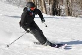
Can I return to jogging, skiing, and other high-impact or contact sports?
If you comply with your physical therapy and post-operative instructions, achieving total muscle recovery, then there are no restrictions on any type of activity. However, studies have shown that, because the implants are mechanical devices, high loads cause them to wear out more quickly, resulting in a higher risk of needing a revision. Please see Your Surgery and Physical Therapy for more information on this condition.
What is TTT?
A tibial tubercle transfer (also known as a Fulkerson Osteotomy) is a surgical procedure that is used to correct for patellar instability or patellar malalignment. Another indication for a tibial tubercle transfer is patellar osteoarthritis. Depending on what anatomy needs to be addressed and corrected, there are a couple choices for repositioning. This will be determined through the use of physical examination and MRI calculations.
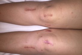
The procedure consists of an incision, which is made a few centimeters below the kneecap (patella) along the top portion of the shin bone (tibia). The patella is embedded in a tendon that inserts on a bony prominence at the shin bone, known as the tibial tuberosity. The patella is repositioned by surgically cutting and moving the attachment on the shin bone. The new position is held through the use of 2 metal screws. The incision is then closed with absorbable sutures and Dermabond, a surgical glue.
How long will I stay in the hospital?
Most patients can go home the same day as their surgery. Occasionally, you will stay overnight. This will allow for better management of your pain. Once you are able to demonstrate successful management of pain, you will be discharged home.
What are the possible risks and complications of surgery?
As with any surgery there is a risk of DVT (blood clot), nerve damage, and postoperative infection. Specific risks and complications include but aren’t limited to failure to heal, fracture, and hardware complications.
When can I drive?
You may not drive while taking pain medication. In addition, if it is your right knee that had surgery, you will not be able to drive for approximately 6 weeks after surgery or until the brace is removed.
When can I resume jogging?
You will not resume jogging until cleared by your physician. This typically is around 6 months after surgery. You will be allowed to begin biking without resistance once you have adequate range of motion and will begin this with your physical therapist. Once adequate quad strength is demonstrated you will transition to the Elliptical, then running is the next step after that.
When can I return to my sport?
There are many factors in returning to sport after surgery. Most patients are able to return around 7-10 months after surgery. Please see Your Surgery and Physical Therapy for more information on this condition.
What makes me a candidate for this surgery?
You are a candidate if you have patellar instability (kneecap dislocation) and front knee pain. This is evaluated through your past history and the use of MRI and XRAY.
What is the medial patellofemoral ligament (MPFL) and how is it damaged?
The MPFL is a ligament that stabilizes the patella (kneecap) and keeps it from displacing laterally. It is disrupted when someone dislocates his/her patella. When the kneecap dislocates towards the outside, this stretches the ligament on the inside of the knee, which is trying to keep the kneecap in place. This can result in either a tear of the MPFL or a detachment of the ligament from the bone.
What does the surgery entail?
A tibial tubercle transfer (also known as a Fulkerson Osteotomy) is a surgical procedure that is used to correct for patellar instability or patellar malalignment.
The procedure consists of an incision, which is made a few centimeters below the kneecap (patella) along the top portion of the shin bone (tibia). The patella is embedded in a tendon that inserts on a bony prominence at the shin bone, known as the tibial tuberosity. The patella is repositioned by surgically cutting and moving the attachment on the shin bone. The new position is held through the use of 2 metal screws. Then, the injured MPFL ligament will be replaced with a graft, usually a hamstring tendon from the same leg or a cadaver allograft. The graft is attached to the patella via small absorbable screws that hold the graft in place. The incision is then closed with absorbable sutures and Dermabond, a surgical glue and tape.
How long will I stay in the hospital?
With this surgery, you will stay overnight. This will allow for better management of your pain. Once you are able to demonstrate successful management of pain, you will be discharged home.
What are the possible risks and complications of surgery?
As with any surgery there is a risk of DVT, nerve damage, and postoperative infection. Specific risks and complications include but aren’t limited to failure to heal, fracture, and hardware complications.
When can I drive?
You may not drive while taking pain medication. In addition, if it is your right knee that had surgery, you will not be able to drive for approximately 6 weeks after surgery or until the brace is removed.
When can I resume jogging?
You will not resume jogging until cleared by your physician. This typically is around 8-10 months after surgery. You will be allowed to begin biking without resistance once you have adequate range of motion and will begin this with your physical therapist. Once adequate quad strength is demonstrated you will transition to the Elliptical, then running is the next step after that.
When can I return to my sport?
There are many factors in returning to sport after surgery. Most patients are able to return around 7-10 months after surgery. Please see Your Surgery and Physical Therapy for more information on this condition.
What makes me a candidate for this surgery?
You are a candidate if you have patellar instability (kneecap dislocation), front knee pain, and focal cartilage injury. This is evaluated through your past history and the use of MRI and XRAY.
What is the medial patellofemoral ligament (MPFL) and how is it damaged?
The MPFL is a ligament that stabilizes the patella (kneecap) and keeps it from displacing laterally. It is disrupted when someone dislocates his/her patella. When the kneecap dislocates towards the outside, this stretches the ligament on the inside of the knee, which is trying to keep the kneecap in place. This can result in either a tear of the MPFL or a detachment of the ligament from the bone.
What is cartilage, why is it important, and how is it injured?
Cartilage is the shiny, smooth coating at the end of bones. It protects the bone and allows the bones to move smoothly and efficiently. Damaged cartilage is known as arthritis. When cartilage thins, or has a piece missing, it puts more stress on the bone and causes pain. The mechanism of injury for a cartilage defect is usually related to trauma, such as a dislocation that causes the cartilage to scrape along bone, or chronic friction from a maligned knee. The body cannot regenerate this type of cartilage.
What does the surgery entail?
A tibial tubercle transfer (also known as a Fulkerson Osteotomy) is a surgical procedure that is used to correct for patellar instability or patellar malalignment. The procedure consists of an incision, which is made a few centimeters below the kneecap (patella) along the top portion of the shin bone (tibia). The patella is embedded in a tendon that inserts on a bony prominence at the shin bone, known as the tibial tuberosity. The patella is repositioned by surgically cutting and moving the attachment on the shin bone. The new position is held through the use of 2 metal screws.
Then, the injured MPFL ligament will be replaced with a graft, usually a hamstring tendon from the same leg or a cadaver allograft. The graft is attached to the patella via small absorbable screws that hold the graft in place.
Using arthroscopy, the cartilage defect is debrided and restored using one of the methods described below.
The incision is then closed with absorbable sutures and Dermabond, a surgical glue and tape.
How will my cartilage be repaired?
This depends on many factors including your age as well as the size and location of the cartilage defect. The surgeon will chose from one of the following procedures:
– Microfracture
- Procedure: Using arthroscopy, the surgeon will first remove any defective and damaged cartilage tissue from the knee joint. She will then create small holes at the site of your lesion to allow bleeding which will stimulate healing. The new tissue that grows is a hybrid of fibrocartilage and another type of cartilage that is similar to that originally in the joint. Although it is not exactly the same, this new type of cartilage is shown to be durable and to function similarly to the original articular cartilage. This procedure can be used on very small lesions successfully.
– DeNovo
- Procedure: The surgeon will first remove any defective and damaged cartilage tissue from the knee joint. She will then patch the cartilage defect with the DeNovo cartilage cells. This consists of juvenile cartilage which is able to rejuvenate and grow.
- You may require follow-up MRIs at 3 mo, 6 mo, 12 mo, and 24 mo after surgery to follow the growth and maturation of the new cartilage.
– OCA (Osteochondral Allograft)
- Procedure: The surgeon will first remove any defective and damaged cartilage tissue from the knee joint. She will then patch the cartilage defect with a plug of allograft donor cartilage and bone. This does not rejuvenate, but is press fit into the bone. The body then grows into the new plug and it becomes your own.
– MACI (autologous cultured chondrocytes on porcine collagen membrane)
- Procedure: This is a staged surgery. Stage 1- Using arthroscopy, the surgeon will first remove any defective and damaged cartilage tissue from the knee joint. Healthy cartilage is biopsied and sent to a lab where the cartilage cells will proliferate. Stage 2- About 4-6 weeks later, the defect will be patched with your new cartilage cells.
How long will I stay in the hospital?
With this surgery, you will stay overnight. This will allow for better management of your pain. Once you are able to demonstrate successful management of pain, you will be discharged home.
What are the possible risks and complications of surgery?
As with any surgery there is a risk of DVT, nerve damage, and postoperative infection. Specific risks and complications include but aren’t limited to failure to heal, fracture, and hardware complications.
When can I drive?
You may not drive while taking pain medication. In addition, if it is your right knee that had surgery, you will not be able to drive for approximately 6 weeks after surgery or until the brace is removed.
When can I resume jogging?
You will not resume jogging until cleared by your physician. This typically is around 8-10 months after surgery. You will be allowed to begin biking without resistance once you have adequate range of motion and will begin this with your physical therapist. Once adequate quad strength is demonstrated you will transition to the Elliptical, then running is the next step after that.
When can I return to my sport?
There are many factors in returning to sport after surgery. Most patients are able to return around 7-10 months after surgery. Please see Your Surgery and Physical Therapy for more information on this condition.
Patient Stories
Thomas Guardino
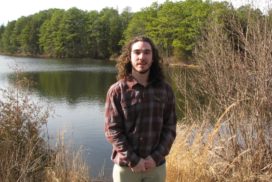
When my experience with Hospital for Special Surgery first began, I was in a poor position mentally and physically, and my outlook on things weren’t the best. I had been competitively playing soccer in the Tri-State area since I was four-years-old. My soccer career included playing competitively in the highest division of the Monmouth and… Read more “Thomas Guardino”
Ava Harrison
When I was in 7th grade I dislocated my knee cap for the very first time. It wasn’t until I was in 8th grade when it dislocated again and my parents and I knew something was wrong with my knees. I didn’t go to HSS right away because we didn’t know if it would be… Read more “Ava Harrison”
Paulette Gangemi
I just wanted to take a moment and share my story and wonderful experience with The Women’s Sports Medicine Center and Doctor Sabrina Strickland at Hospital for Special Surgery. As you can see, I am not “local” to NYC. It is an effort to come to Manhattan, but the effort it well worth it. I… Read more “Paulette Gangemi”
Katherine Trumble
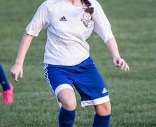
I first injured myself playing basketball when I had just turned eleven years old. I was side shuffling to block a shot when my right knee locked and I fell down, dislocating my patella. I did not know what happened and when I was told I would be out of sports for some time, I… Read more “Katherine Trumble”
Maureen Suhr

Since adolescence, I have had knee pain and subluxation and many knee surgeries. Knee pain was a part of my life for over twenty years, but it became increasingly worse to the point where I had episodes of significant swelling every other week. It began interfering with my work, my commute, and limited my activities… Read more “Maureen Suhr”
Laura Carhart
I am 27 years old. A few years ago my knee cap was on the left side of my knee and half of my cartilage was gone on the right side of my knee. So my doctor down here told me he could not do it and he sent me up to HSS see Dr.… Read more “Laura Carhart”
Seyward Darby

My left kneecap started dislocating and subluxating in early adolescence, when I was playing soccer and basketball regularly and also growing tall rather quickly (I wound up close to 5’8). Over the next 15 years, the condition was a constant in my life–I lived with the pain when my kneecap popped out of place a… Read more “Seyward Darby”
Sarah Perry
I have dealt with knee pain for eight years and finally decided to take the necessary measures to live a pain-free life. I came to HSS with hopes of being able to run, Irish-dance, take yoga, and have zero pain in daily activities. I have been active in various team sports throughout the years, but… Read more “Sarah Perry”
